
January 3, 2017
Lipid metabolism is potential ‘Achilles’ heel’ for cancer stem cells
WEST LAFAYETTE, Ind. — Researchers have discovered a metabolic signature critical for the functioning of “cancer stem cells” that initiate tumor formation. The team also showed how to interfere with this metabolic mechanism in ovarian cancer, inhibiting tumor growth.
“The cancer stem cells are resistant to conventional therapies and are responsible for tumor relapse after chemotherapy and development of distant metastases,” said Ji-Xin Cheng, a professor in Purdue University's Weldon School of Biomedical Engineering and Department of Chemistry. “Understanding their unique characteristics and vulnerabilities will enable the development of targeted therapies with the ultimate goal of overcoming tumor relapse and metastasis.”
New research focuses on targeting cancer stem cells by inhibiting the activities of enzymes needed to carry out a metabolic process called “desaturation” of lipid molecules.
“Unsaturated lipids in the cancer stem cells are very important to maintain the signaling needed to function,” said Cheng, a member of Purdue’s Center for Cancer Research. “Researchers have known about cancer stem cells for a while, but lipid metabolism in these cells is a very new topic. Understanding the lipid metabolism in cancer stem cells opens a new way for cancer detection and treatment.”
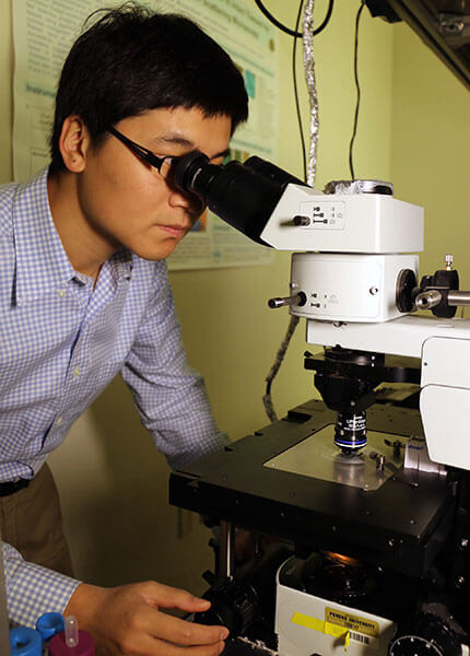 Purdue postdoctoral research associate Junjie Li is a co-first author of a paper appearing in the journal Cell Stem Cell regarding research into “cancer stem cells” that initiate tumor formation. The research is led by Ji-Xin Cheng, a professor in Purdue's Weldon School of Biomedical Engineering and Department of Chemistry. (Purdue University photo/Erin Easterling)
Download image
Purdue postdoctoral research associate Junjie Li is a co-first author of a paper appearing in the journal Cell Stem Cell regarding research into “cancer stem cells” that initiate tumor formation. The research is led by Ji-Xin Cheng, a professor in Purdue's Weldon School of Biomedical Engineering and Department of Chemistry. (Purdue University photo/Erin Easterling)
Download image
The work was performed by researchers at Purdue, Indiana University School of Medicine, and Northwestern University’s Feinberg School of Medicine.
Findings are detailed in a research paper that appeared online Dec. 29 in the journal Cell Stem Cell. The paper will be published in the journal’s March 2 print issue.
“In this study, we identify and characterize for the first time lipid unsaturation in ovarian cancer stem cells by chemical imaging of single living cells,” said Purdue postdoctoral research associate Junjie Li, who was the paper’s co-first author along with Salvatore Condello, a research assistant professor in the Feinberg School of Medicine.
The researchers demonstrated that a specific “signaling pathway” directly regulates the production of lipid enzymes, called fatty acid desaturases.
“Collectively, our findings reveal that increased lipid unsaturation is a metabolic marker for ovarian cancer stem cells and a target for therapy focusing on these types of cells,” said Cheng, working with the Northwestern University team led by Daniela Matei, a professor in the Feinberg School of Medicine.
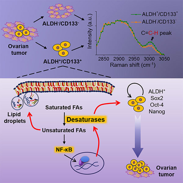 This graphic illustrates an approach to potentially treat ovarian cancer by interfering with the metabolic mechanism of “cancer stem cells,” which initiate tumor formation. (Purdue University photo/Ji-Xin Cheng)
Download image
This graphic illustrates an approach to potentially treat ovarian cancer by interfering with the metabolic mechanism of “cancer stem cells,” which initiate tumor formation. (Purdue University photo/Ji-Xin Cheng)
Download image
Identifying unique metabolic traits, which can be exploited as an Achilles’ heel to eliminate cancer stem cells, is an important step forward, said Matei, co-senior author of the paper.
“Here we propose a completely new strategy to eradicate recalcitrant cancer cells responsible for tumor recurrence after standard treatment and elucidate how lipid metabolism contributes to the survival of ovarian cancer stem cells,” she said.
A YouTube video is available at https://youtu.be/4NoHL1lk7QU
The paper was authored by Li; Condello; gynecologic oncologist Jessica Thomes-Pepin, formerly at IU and now at Minnesota Oncology; former Purdue postdoctoral research associate Xiaoxiao Ma; Yu Xia, an associate professor of chemistry at Purdue; Thomas D. Hurley, a professor in the IU Department of Biochemistry and Molecular Biology; Matei; and Cheng.
Until now, the lack of sensitive single-cell analysis tools has limited the characterization of metabolic activity in cancer stem cells. The imaging methods used in the study were developed by Cheng’s group and allow researchers to detect the signature in single cells. Because cancer stem cells represent a tiny population of the total number of cancer cells, the single-cell sensitivity is important for detecting hidden metabolic signatures.
The new approach uses two technologies: hyperspectral stimulated Raman scattering imaging of single living cells and mass spectrometry analysis of extracted lipids.
“We report here significantly increased levels of unsaturated lipids in ovarian cancer stem cells compared to non-cancer stem cells,” Cheng said. “Conventional methods can’t do single cell analysis. If the signature is localized in a very small area, it is not easily detected by conventional biochemical assay. By stimulated Raman microscopy, we can better pinpoint these cells through the metabolic signature.”
The lipid-desaturation signature was identified in laboratory cultures of ovarian cancer stem cells and in cells from human patients. Higher lipid unsaturation levels also were detected in cancer stem cell-enriched laboratory cultures called “spheroids,” a three-dimensional culture that mimics tumors in human patients. The researchers used a chemical to inhibit the activities of desaturases and reduce the “stemness” of the cells, rendering them less lethal. Inhibition of lipid desaturases effectively eliminated cancer stem cells, suppressed spheroid formation in laboratory cultures, and blocked tumor initiation capacity in laboratory mice, Cheng said.
“We don’t directly block the signaling pathway, but we block fatty acid metabolism so that the unsaturated lipids are reduced, and that actually suppresses the function of the cancer stem cells,” Li said.
Some of the research was done in the Bindley Bioscience Center in Purdue’s Discovery Park and was supported by the Purdue Center for Cancer Research.
The researchers have filed a patent application through the Office of Technology Commercialization of the Purdue Research Foundation.
The research, which is ongoing and may progress into clinical studies, also was supported by the National Institutes of Health and the U.S. Department of Veterans Affairs.
Writer: Emil Venere, 765-494-4709, venere@purdue.edu
Source: Ji-Xin Cheng, 765-494-4335, jcheng@purdue.edu
Note to Journalists: An electronic copy of the research paper is available by contacting Emil Venere, 765-494-4709, venere@purdue.edu, or the Cell Press, press@cell.com, 617-397-2802. A YouTube video is available at https://youtu.be/4NoHL1lk7QU, and other materials are available on Google drive at https://goo.gl/FSUjtO. The materials were prepared by Erin Easterling, digital producer in Purdue’s College of Engineering.
ABSTRACT
Lipid Desaturation Is a Metabolic Marker and Therapeutic Target of Ovarian Cancer Stem Cells
Junjie Li1*, Salvatore Condello2*, Jessica Thomes-Pepin3, Xiaoxiao Ma4, Yu Xia4, Thomas D. Hurley5, 6, Daniela Matei2,7,8#, Ji-Xin Cheng1,4,9,10#
1 Weldon School of Biomedical Engineering, Purdue University, West Lafayette, IN 47907
2 Department of Obstetrics and Gynecology, Feinberg School of Medicine, Northwestern University, Chicago, IL, 60611
3 Department of Obstetrics and Gynecology, Indiana University, Indianapolis, IN 46202
4 Department of Chemistry, Purdue University, West Lafayette, IN 47907
5 Department of Biochemistry and Molecular Biology, Indiana University School of Medicine, Indianapolis, IN 46202
6 Indiana University Simon Cancer Center, Indiana University School of Medicine, Indianapolis, IN 46202
7 Jesse Brown VA Medical Center, Chicago, IL, 60611
8 Robert H. Lurie Comprehensive Cancer Center, Chicago, IL, 60611
9 Purdue University Center for Cancer Research, Purdue University, West Lafayette, 47907
10 Lead contact, * Co-first author, # Correspondence: daniela.matei@northwestern.edu (D.M.), jcheng@purdue.edu (J.X.C)
Lack of sensitive single-cell analysis tools has limited the characterization of metabolic activity in cancer stem cells. By hyperspectral stimulated Raman scattering imaging of single living cells and mass spectrometry analysis of extracted lipids, we report here significantly increased levels of unsaturated lipids in ovarian cancer stem cells (CSCs) as compared to non-CSCs. Higher lipid unsaturation levels were also detected in CSC-enriched spheroids compared to monolayer cultures of ovarian cancer cell lines or primary cells. Inhibition of lipid desaturases effectively eliminated CSCs, suppressed sphere formation in vitro, and blocked tumor initiation capacity in vivo. Mechanistically, we demonstrate that NF-κB directly regulates the expression levels of lipid desaturases and that inhibition of desaturases blocks NF-κB signaling. Collectively, our findings reveal that increased lipid unsaturation is a metabolic marker for ovarian CSCs and a target for CSC-specific therapy.
