February 28, 2018
3-D-written model to provide better understanding of cancer spread
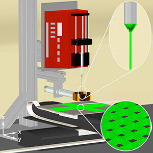
 This diagram conceptualizes the 3-D jet writer a Purdue researcher is using to engineer a cancer microenvironment. (University of Michigan image/Jacob Jordahl)
Download image
WEST LAFAYETTE, Ind. — Purdue researcher Luis Solorio has helped create a lifelike cancer environment out of polymer to better predict how drugs might stop its course.
Previous research has shown that most cancer deaths happen because of how it spreads, or metastasizes, in the body. A major hurdle for treating cancer is not being able to experiment with metastasis itself and knock out what it needs to spread.
Studies in the past have used a 3-D printer to recreate a controlled cancer environment, but these replicas are still not realistic enough for drug screening.
“We need a much finer resolution than what a 3-D printer can create,” said Solorio, an assistant professor of biomedical engineering.
Rather than 3-D printing, Solorio and a team of researchers have proposed 3-D writing. The device that they developed, a 3-D jet writer, acts like a 3-D printer by producing polymer microtissues as they are shaped in the body, but on a smaller, more authentic scale with pore sizes large enough for cells to enter the polymer structure just as they would a system in the body.
3-D jet writing is a fine-tuned form of electrospinning, the process of using a charged syringe containing a polymer solution to draw out a fiber, and then deposit the fiber onto a plate to form a structure. This structure is a scaffold that facilitates cell activity.
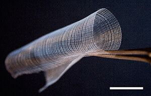
 A 3-D jet writer produces polymer structures that model the biological tissue cancer cells penetrate. (University of Michigan image/Jacob Jordahl)
Download image
Solorio has so far used the device to write a structure that drew in cancer cells to sites in mice where cancer would not normally develop, confirming that the device could create a feasible cancer environment. Solorio’s other studies have increased cancer cells in human samples for better analysis and maintained receptors on these cells that drugs would need to find.
“Ideally, we could use our system as an unbiased drug screening platform where we could screen thousands of compounds, hopefully get data within a week, and get it back to a clinician so that it’s all within a relevant time frame,” Solorio said.
Initial findings published on Feb. 27 in Advanced Materials based on Solorio’s work as part of a team at the University of Michigan Biointerfaces Institute. Continuation of the work is being performed at Purdue and is funded by the National Cancer Institute grant R0019829.
Writer: Kayla Wiles, 765-494-2432, wiles5@purdue.edu
Source: Luis Solorio, 765-496-1956, lsolorio@purdue.edu
ABSTRACT
3D Jet Writing: Functional microtissues based on tessellated scaffold architectures
The advent of adaptive manufacturing techniques supports the vision of cell-instructive materials that mimic biological tissues. 3D jet writing, a modified electrospinning process reported herein, yields 3D structures with unprecedented precision and resolution offering customizable pore geometries and scalability to over tens of centimeters. These scaffolds support the 3D expansion and differentiation of human mesenchymal stem cells in vitro. Implantation of these constructs leads to the healing of critical bone defects in vivo without exogenous growth factors. When applied as a metastatic target site in mice, circulating cancer cells home in to the osteogenic environment simulated on 3D jet writing scaffolds, despite implantation in an anatomically abnormal site. Through 3D jet writing, the formation of tessellated microtissues is demonstrated, which serve as a versatile 3D cell culture platform in a range of biomedical applications including regenerative medicine, cancer biology, and stem cell biotechnology.

