
| RELATED INFO |
| * Bindley Bioscience Center |
| * Birck Nanotechnology Center |
| * Purdue School of Veterinary Medicine |
| * South Korean Ministry of Science and Technology |
| * Discovery Park |
![]()
![]()
July 10, 2007
Purdue, S. Korean researchers collaborate on nanomedicine project
WEST LAFAYETTE, Ind. -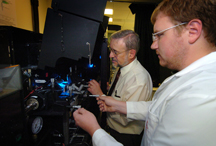 |
The Korean Institute of Science and Technology, also known as KIST, and Purdue developed a research proposal utilizing the complementary strengths of both institutions' internationally renowned research groups.
The South Korean Ministry of Science and Technology selected the KIST and Purdue team project from 20 international research proposals submitted to examine how to learn more about the molecular makeup of diseases. The research initiative, which spans nine years, initially will involve dozens of KIST and Purdue researchers.
 |
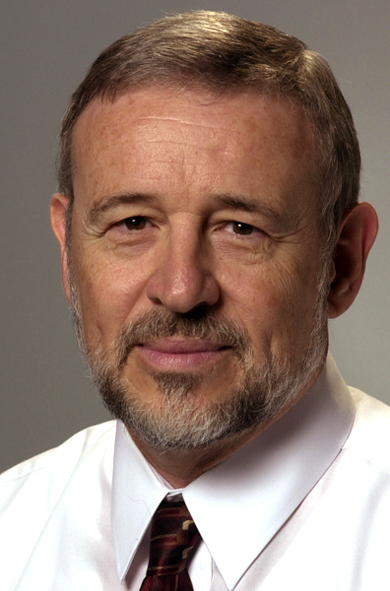
"Theragnosis is a new concept in next-generation medicine that combines simultaneous diagnostics and therapeutics," said Purdue project leader James Leary, the School of Veterinary Medicine Professor of Nanomedicine at Discovery Park's Birck Nanotechnology Center and a professor of basic medical sciences and biomedical engineering.
"In this case, we are combining new molecular imaging, biosensing and nanomedical techniques, linking our state-of-the-art facilities and equipment in Discovery Park with researchers and advanced labs in South Korea. It's truly an international, interdisciplinary team effort."
Joining Leary from Purdue on the project are:
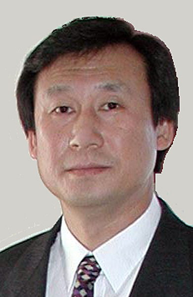
* Kinam Park, the Showalter Distinguished Professor of Biomedical Engineering and a professor of pharmaceutics.
* J. Paul Robinson, professor in the schools of Biomedical Engineering and Veterinary Medicine and director of Purdue's Cytometry Laboratories at the Bindley Bioscience Center.
* Ji-Xin Cheng, an assistant professor in the Weldon School of Biomedical Engineering and Department of Chemistry.
The Purdue team is working with researchers Kuiwon Choi of KIST, who will lead the South Korean research team along with Ick Chan Kwon, the co-principal investigator of the program.
"The collaboration between KIST's Biomedical Research Center and Purdue's Bindley Bioscience Center will be the most promising implement for theragnosis," said Choi, who leads the Biomedical Research Center at KIST and is currently president of the Korean Society of Biomechanics.
As a part of the project, KIST and Purdue researchers will work to advance the role that nanoparticles can play in the diagnosis and treatment of cancer or chronic diseases like diabetes and multiple sclerosis.
"This research program is believed to be the first in the world to integrate molecular imaging and nanomedicine with chemistry, drug delivery, cell biology and molecular biology, as well as cell engineering and clinical medicine," Leary said. "These disciplines together are far more powerful in advancing medicine than they are individually. This advances our research efforts to examine how we can treat diseases at the molecular level."
Leary's team has made advancements in studying how nanoparticles can be used as a new type of therapy and how they can be directed as drug-delivery capsules to seek out and penetrate individual diseased cells to repair or destroy severely damaged cells.
An injection with a hypodermic needle can release millions of these capsules into a person's bloodstream, Leary said. Once there, nanoparticles take advantage of the body's natural cellular signaling system to find the damaged cells.
"Using complex molecules embedded in their outer membranes, the trillions of cells in a human body can identify themselves and communicate with each other," Leary said. "The molecules act as chemical 'flags' for communicating with other cells or as 'gates' that control entrance to the cell for molecules in the bloodstream."
Leary designed a technique for creating a nanoparticle with layers of therapeutic molecules. The layers peel back and specific molecules are activated as they are needed. This bypasses the difficulty of creating a "silver bullet," or a single molecule to solve everything.
By embedding biosensors within the nanoparticle that react to specific biomarkers for disease, the nanoparticles can effectively switch the treatment on or off. This feedback loop enables the therapy to deliver the exact dose of medicine needed and to treat only the diseased cells.
"We are taking medical treatment to the individual cell level and even to the molecules within a cell," Leary said. "That is where the disease is. A nanoparticle could find a cell infected with a virus and disassemble the virus without harming the host cell. Current methods, such as surgery and radiation treatment, are blunt instruments compared to the future of nanomedicine. Through nanomedicine, we could repair infected cells instead of killing them."
 |
For Park, this project advances his research in using various synthetic polymers, such as polymer micelles and hydrogels, for controlled delivery of various molecular imaging systems.
"The emerging field of nanomedicine, specifically employing nanomaterials for in-vivo imaging, diagnostics and therapeutics, is of primary interest for activities at the Purdue Discovery Park and the Bindley Bioscience and Birck Nanotechnology centers," Park said. "International collaborations of this magnitude are crucial to create an environment for interdisciplinary collaborations leading to significant advancements in this arena."
Robinson will employ advancements he has made in his cytometry lab at Bindley that enables the KIST-Purdue team to study as many as 32 different colored tags identifying molecules of interest from a single cell flowing past a laser beam. These tags will yield a wealth of data about particular cells.
 |
In flow cytometry, cells suspended in a liquid are treated with fluorescent dyes called "markers" and made to flow past a laser beam at approximately 10 meters per second. Different markers automatically bind to specific cells, and the colored dyes glow when exposed to the laser beam. Analyzing a single particle or cell in 32 separate colors provides a "spectral signature" that enables researchers to diagnose disease or detect biological and chemical agents, Robinson said.
"With this technique, we can make a full fluorescence profile of a single cell in that brief period of time to help us define various properties of a cell," he said. "Different diseases will be reflected by different proportions of certain types of cells, helping us simultaneously diagnose and treat diseases ranging from anemia to AIDS."
The Purdue team also is studying techniques for attaching fluorescent molecules to nanoparticles to diagnose and treat illnesses. These are designed to light up at certain stages of the process, with some able to employ different colors at different stages. Within a layered nanoparticle, color change could indicate the activity of each layer and therapeutic molecule. They also become tracking devices for the nanoparticles within the human body.
 |
Cheng strengthens the team because of his research in the use of chemically selective microscopic imaging techniques to analyze living tissue and learn more about the molecular mechanisms of chronic diseases and cancer.
Raman microscopy, an imaging technique invented more than three decades ago, cannot be used effectively to study living tissue because the extremely weak "Raman scattering" signals require hours to yield an image, whereas "coherent anti-Stokes Raman scattering," or CARS, overcomes this limitation, Cheng said.
Conventional microscopic imaging techniques require samples to be labeled with dyes, killing the tissues in the process, Cheng said. All molecules within a living cell vibrate, and the vibrations are unique for each type of molecule. By analyzing these vibrations, a researcher can know exactly what molecules are present and their precise location.
Because his imaging techniques work without using dyes to "label" cells and structures, they can be used to study living tissues without altering them, representing a major advantage over conventional microscopic imaging technologies, Cheng said.
"CARS microscopy permits label-free imaging of specific molecules with a speed of one frame per second or even faster," Cheng said. "Because this technique spares us the need to apply labels, we can examine and track these molecules more precisely in their native state, and that's profound in helping us diagnose and treat diseased or damaged cells."
In conjunction with this project, Purdue's Discovery Park will hold an international conference on Sept. 14 in the Burton D. Morgan Center for Entrepreneurship. More than 20 Korean researchers on this project will attend, along with other international scholars in the growing field of molecular imaging and theragnosis. The first event took place in Seoul in September 2006.
"We're starting this partnership with a core group of researchers from KIST and Purdue, but we will seek the full participation of other researchers at Purdue University to jump-start this project and other potential international collaborations," Park said.
The Ministry of Science and Technology, which was launched in 1967, provides central planning, coordination and evaluation of all science and technology activities in South Korea. The agency also formulates national policies in the areas of technology, space and nuclear energy.
Launched in 2001, Purdue's Discovery Park has grown into a $350 million interdisciplinary hub for research, bringing together more than 1,000 Purdue faculty and 2,500 university students to tackle solutions in areas ranging from health care, nanotechnology and life sciences to alternative energy, the environment and climate change.
Writers: Phillip Fiorini, (765) 496-3133, pfiorini@purdue.edu
Elizabeth K. Gardner, (765) 494-2081, ekgardner@purdue.edu
Sources: James Leary, (765) 494-7280, jfleary@purdue.edu
Kinam Park, (765) 463-1989, kpark@purdue.edu
J. Paul Robinson, (765) 494-0757, jpr@flowcyt.cyto.purdue.edu
Ji-Xin Cheng, (765) 494-4335, jcheng@purdue.edu
Kuiwon Choi, choi@kist.re.kr
Purdue News Service: (765) 494-2096; purduenews@purdue.edu
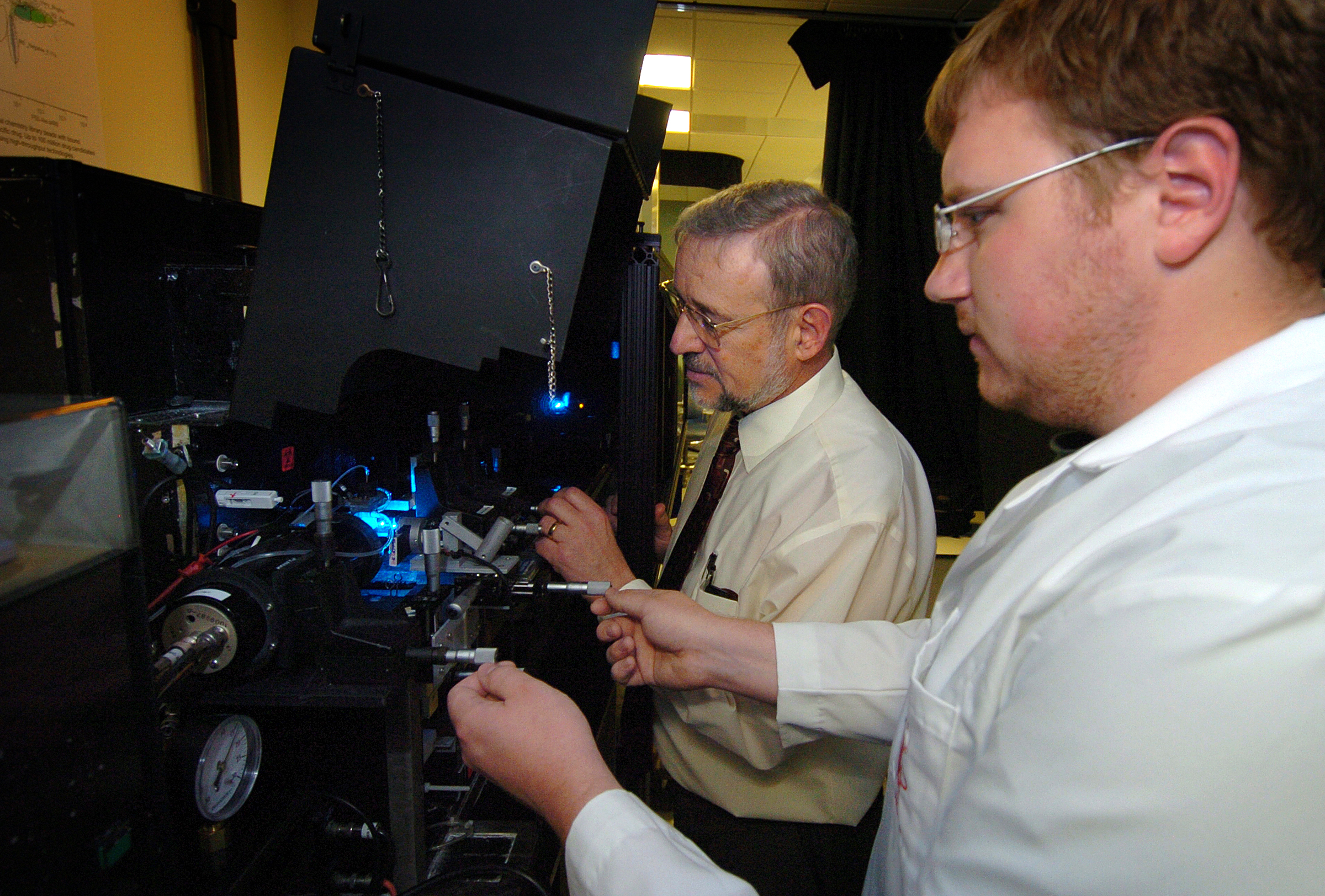
PHOTO CAPTION:
James Leary, from left, a School of Veterinary Medicine professor of nanomedicine and professor of biomedical engineering at Purdue, and Michael Zordan, a graduate student in biomedical engineering, prepare samples for a special high-speed cell sorter at Discovery Park's Bindley Bioscience Center. This instrument, located in Bindley's Molecular Cytometry Laboratory, is a key part of a $4.5 million project that combines molecular imaging, cytometry and nanomedicine to diagnose and treat illnesses and diseases at the molecular level. (Purdue News Service photo/David Umberger)
A publication-quality photo is available at https://www.purdue.edu/uns/images/+2007/leary-cellsorter.jpg
To the News Service home page
![]()