Lab Culture: Trask Lab
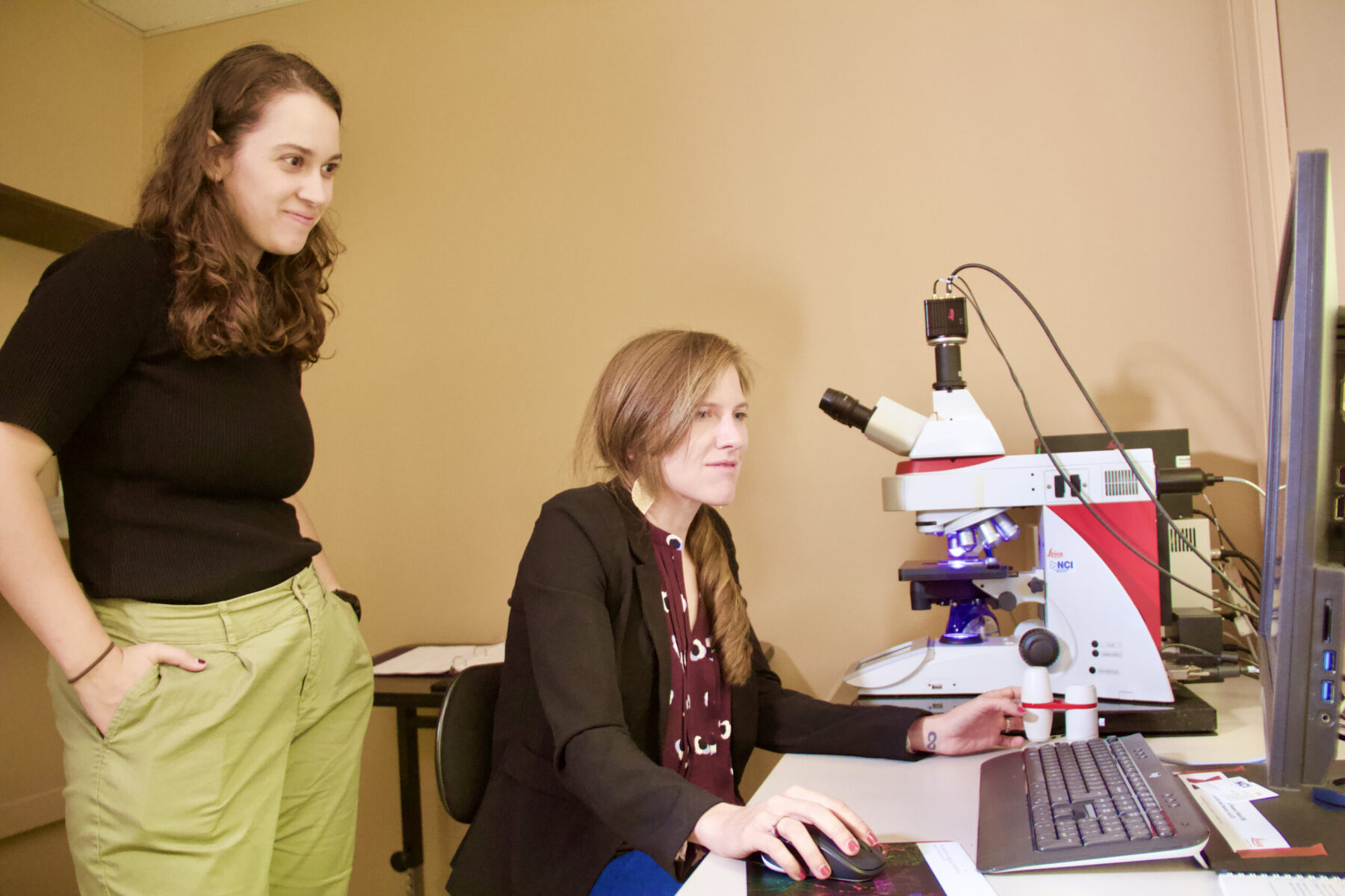
Psychological sciences Assistant Professor Sydney Trask sits at her fluorescence microscope while graduate student Erisa Met Hoxha observes.Tim Brouk
Written by: Tim Brouk, tbrouk@purdue.edu
While many scientists who study neurodegeneration focus on the hippocampus area in the brain, Sydney Trask, Purdue University assistant professor in the College of Health and Human Sciences’ Department of Psychological Sciences, looks at the retrosplenial cortex because of the memories stored within. This work aims to discover new pathways in the understanding of age-related neurodegenerative conditions such as dementia.
“It seems to be the place where memory is stored more permanently,” Trask said. “The hippocampus is important for memory specificity, but your memory becomes less specific as time goes on. You can tell me what happened yesterday, but what about what happened three weeks ago? It’s a little more fuzzy.
“The retrosplenial holds on to information longer term. … If I ask you about something three years ago, your retrosplenial is going to be important, and your hippocampus won’t be important at all.”
Her research often measures levels of zif268 protein in the retrosplenial cortex. Trask said high levels of the protein are among the first signs of the onset of disregulated memory in old age. Protein is cleared away in the brain when a memory is retrieved, Trask said. When memories fade, the protein is not cleared as fast due to the brain’s molecular ubiquitin-proteasome system’s (UPS) failure.
“There’s a disengagement of this UPS that causes protein accumulation,” she continued. “This coincides with impairments in memory retention. They just don’t learn as well, and they don’t remember what they learned as well.”
Pavlovian influences
Psychology 101 will teach you about the mental conditioning work of Ivan Pavlov and his many dogs. The early 20th century Russian scientist exposed the canines to sounds like bells and metronomes during feeding times. He then measured the dogs’ responses in drool measurement when they heard those sounds when it wasn’t time to eat.
Trask utilizes tried-and true-Pavlovian methodology in her lab. She measures the brains of her rat models after conditioning experiments. This is done through a sophisticated camera system capturing her rats’ behaviors by changing their environments.
“What we’re typically looking for is how patterns of activity change following distinct learning events,” Trask said. “So, when animals are learning about environment A or environment B based on their prior history with those environments, what’s really going on in the brain, and what’s changing in response to that?”
Sweet ’scope
Trask’s lab consists of several rooms in the Psychological Sciences Building. One space is dominated by a powerful Leica fluorescence microscope that can not only enlarge brain tissue to extreme levels, but it can also look at multiple samples at a time. This kind of microscope uses a mercury or xenon lamp to produce ultraviolet light, and it is commonly used in observing molecules within cells and cells within tissues. A slide may have 10 models on it, and Trask is able to see everything at once on a large computer screen. She can click around and zoom in much like Google Earth.
“This microscope is interesting because it allows us to take a wide-field view of exactly where we are in the brain. When I click, the microscope will go there itself and take a fine-grained detailed picture at something like 40x or 20x,” Trask explained. “You can see distinct patterns of activity throughout this brain by taking pictures of it zoomed in really, really far — about 200 times of what you’d normally see. We can look at distinct populations of cells and their neural activity associated with each event that we’re interested in studying.”
The microscope has been huge for the researcher, who began her Purdue career last August.
“We can take these pictures concurrently so that we’re getting a whole bunch of data all at once,” Trask said. “This allows us to map the entire brain in a way that on previous microscopes and other typical fluorescent ’scopes would have taken months to do. I can do it in a matter of days here.”
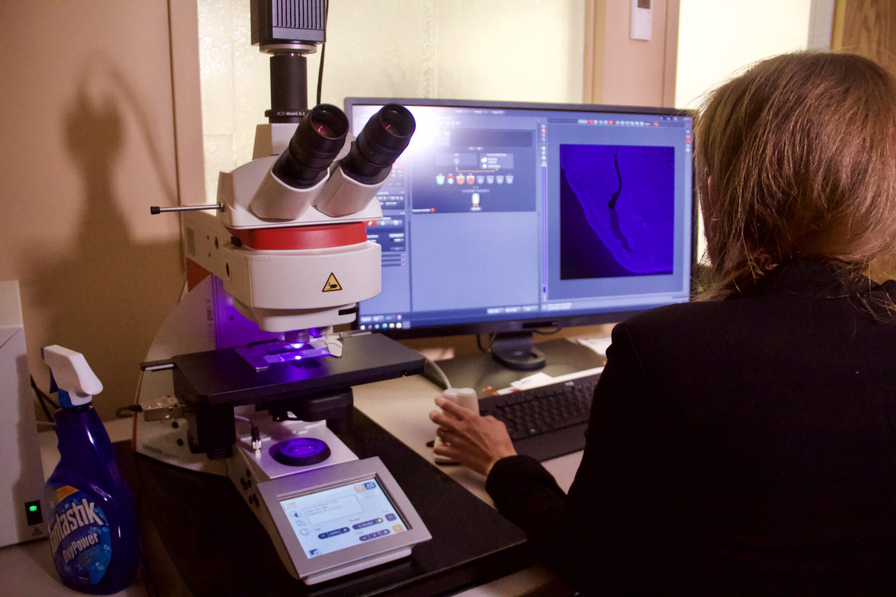
Trask can zoom in on a specimen by 200 times with a simple mouse click.Tim Brouk
Discover more from News | College of Health and Human Sciences
Subscribe to get the latest posts to your email.